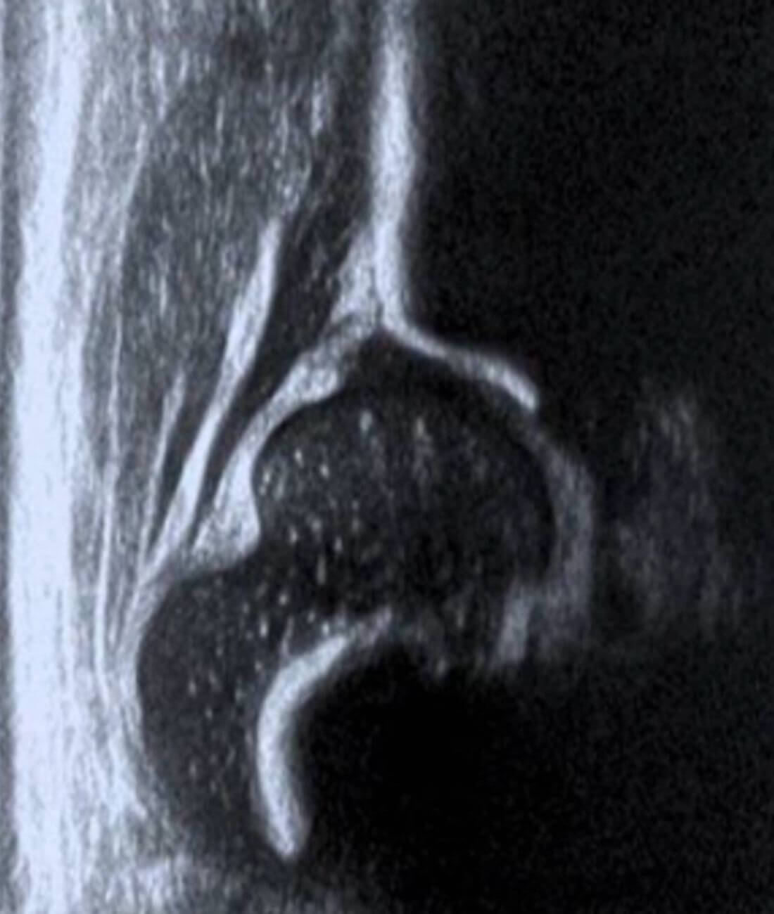
A standardized ultrasound diagnostic method for the early and timely detection of hip dysplasia. This method is based on the use of an ultrasound image in a static coronal view of the hip, where measurements of both the alpha angle (bony acetabular coverage) and the beta angle (coverage from the cartilaginous roof of the hip) should be considered, determining whether the hip in question is centered or not centered; and classifying the hips into 4 possible groups, with their respective subdivisions, thus guiding timely treatment and follow-up depending on the ultrasound findings.
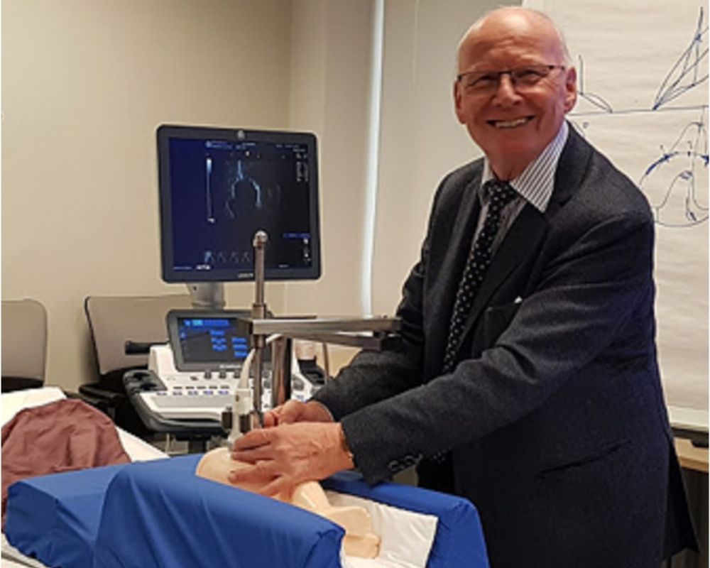
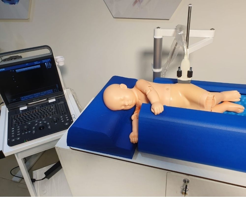
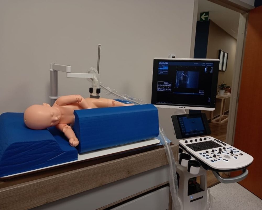
To perform the study using the Graf method, a standardized position for both the baby and the ultrasound machine is essential. The use of the crib and mechanical arm designed by Professor Graf is indispensable, as it reduces bias and human error during the examination. This standardized position specifically requires placing the thumb and index fingers on the greater trochanter. The transducer will then be placed between these fingers (which remain pinched) and always parallel to the edge of the crib.
Simple Scanning Technique
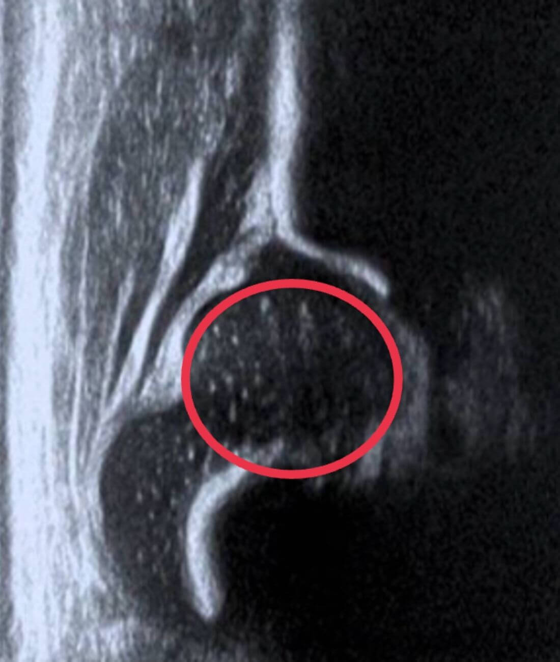
The transducer is moved back and forth over the greater trochanter. Emphasis will be placed on finding the most ossified part of the ilium, referred to as the lower limb.
Correction of the plane by performing a slight rotation of the transducer guide.
Two checklists must always be sought. These will be crucial for validating each image.
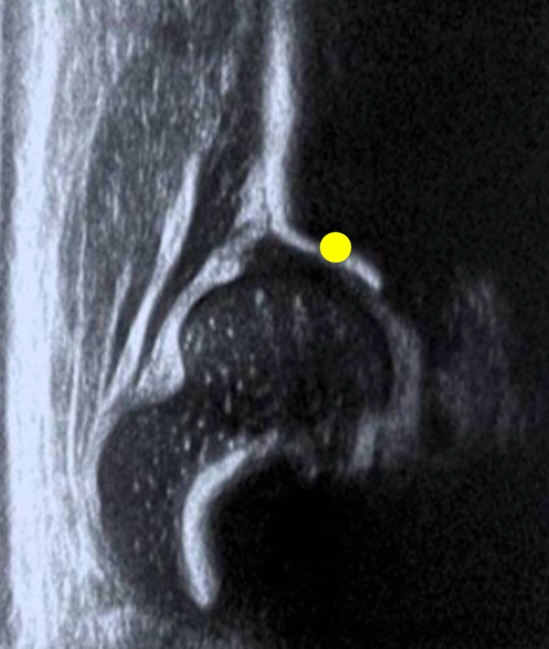
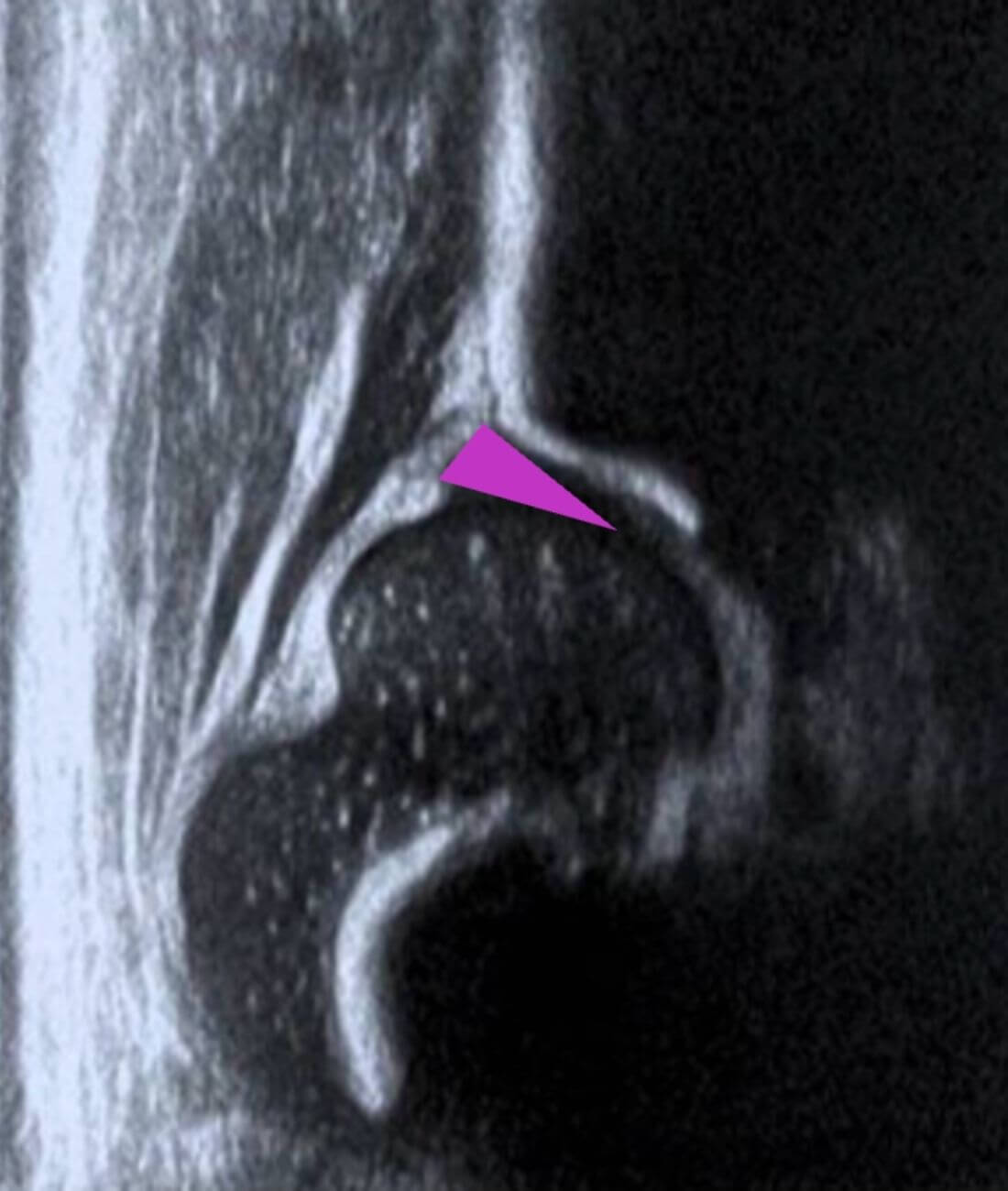
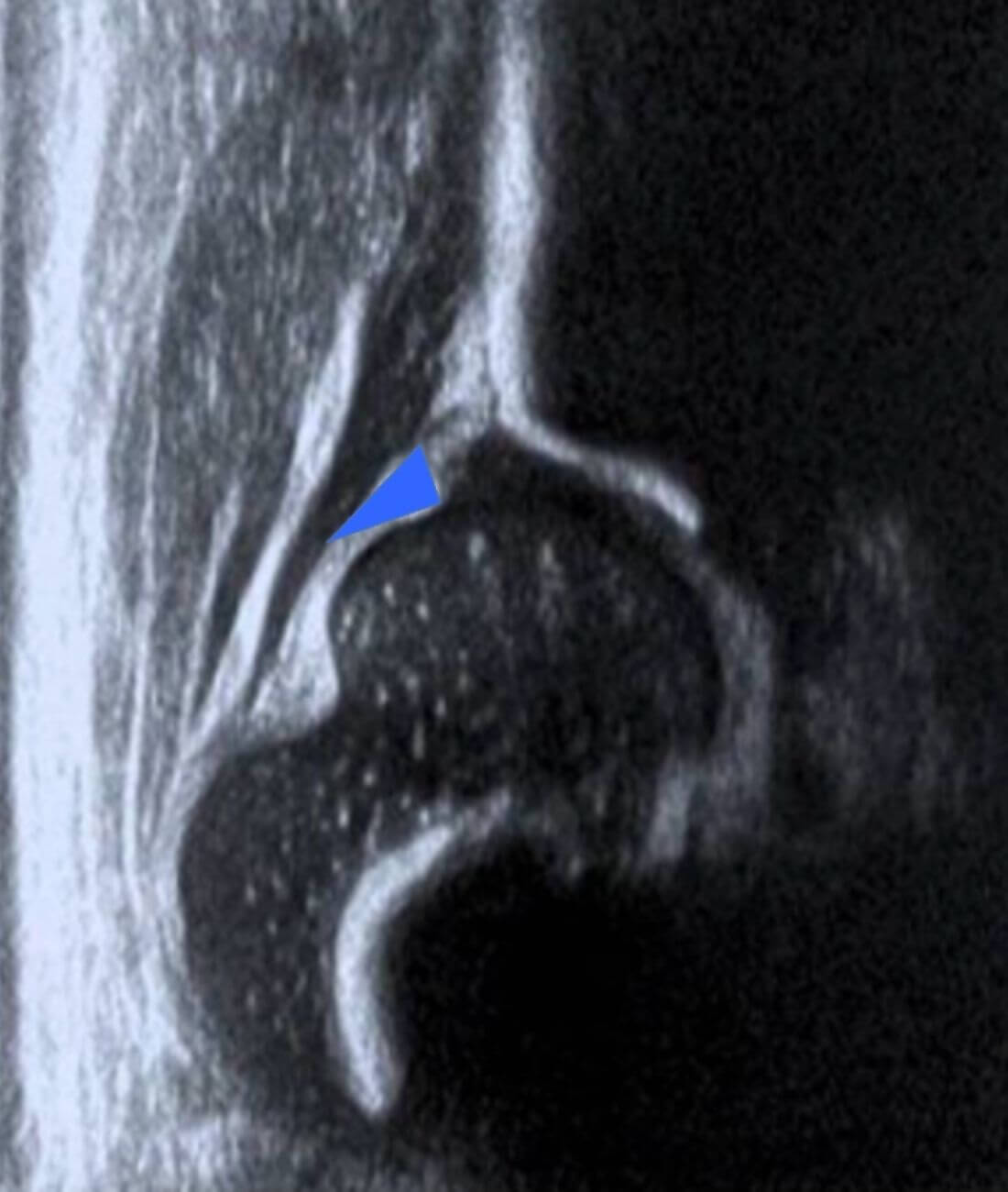
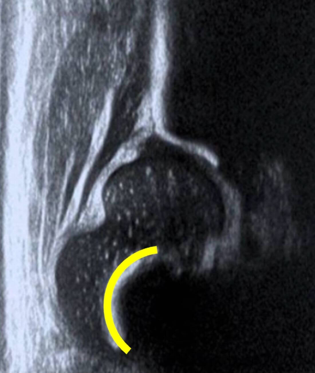
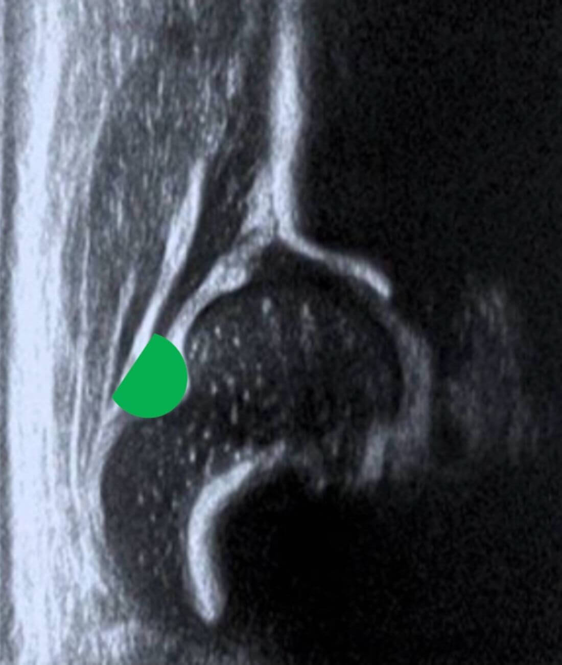
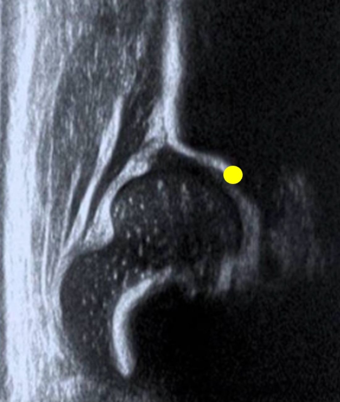
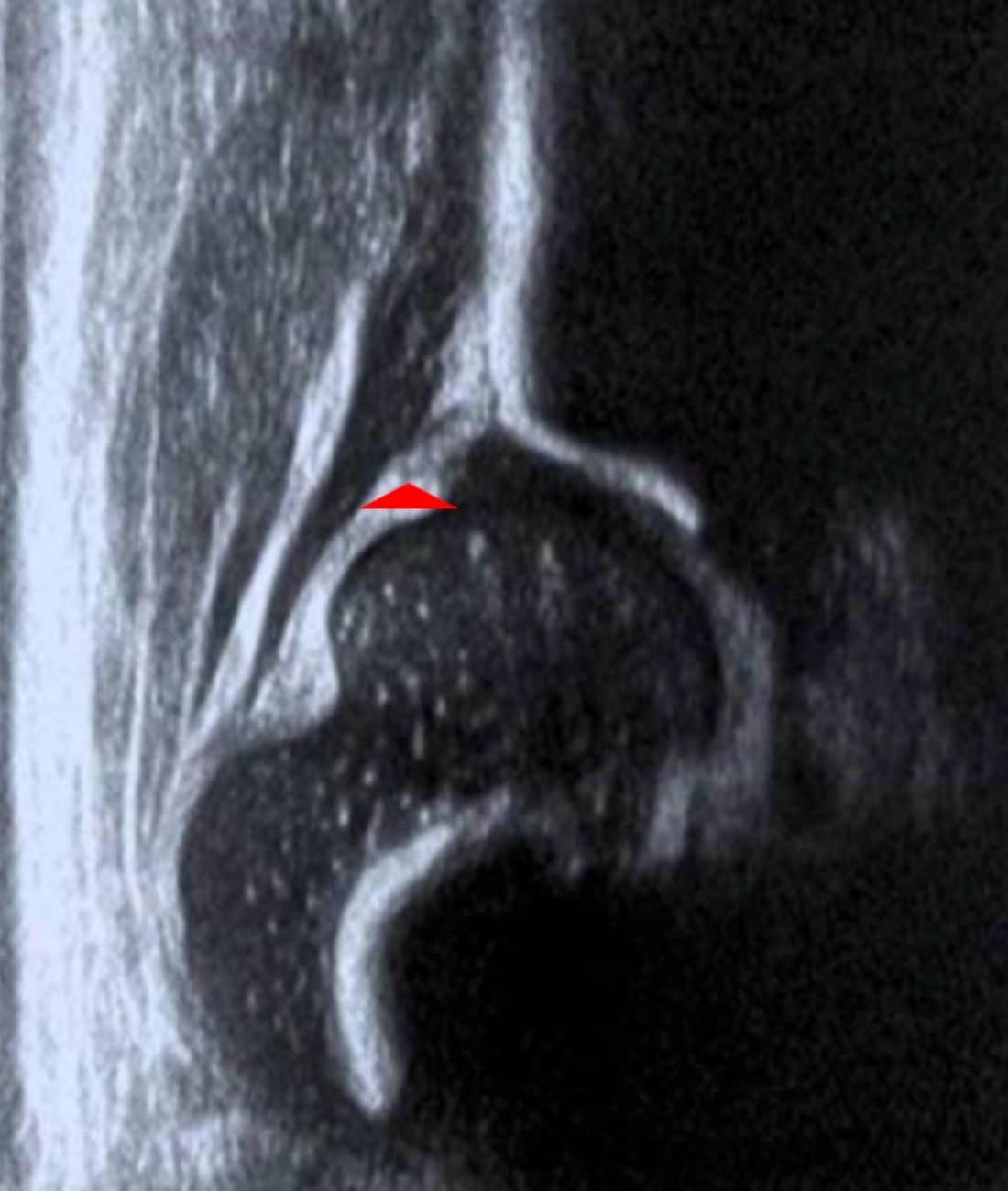
The first checklist is known as the anatomical checklist and should show 7 anatomical structures as well as the turning point.
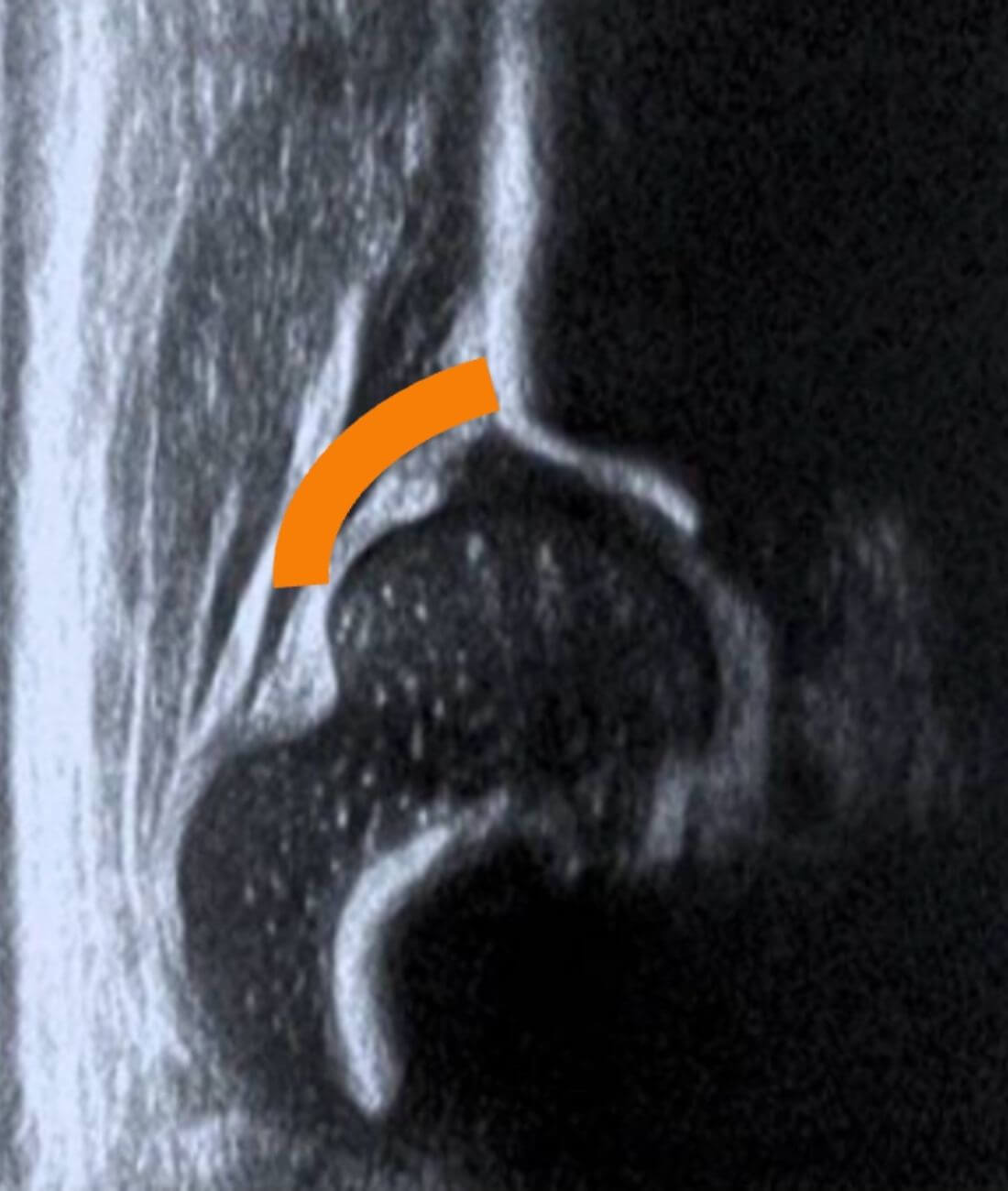
The second checklist will show 3 structures that are essential for validating the standardized position of the image.
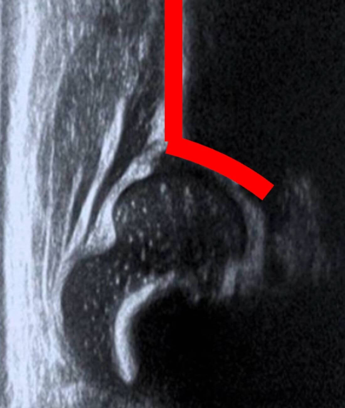
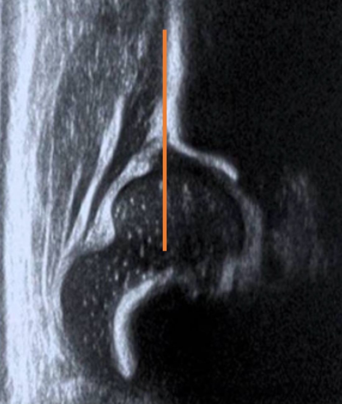
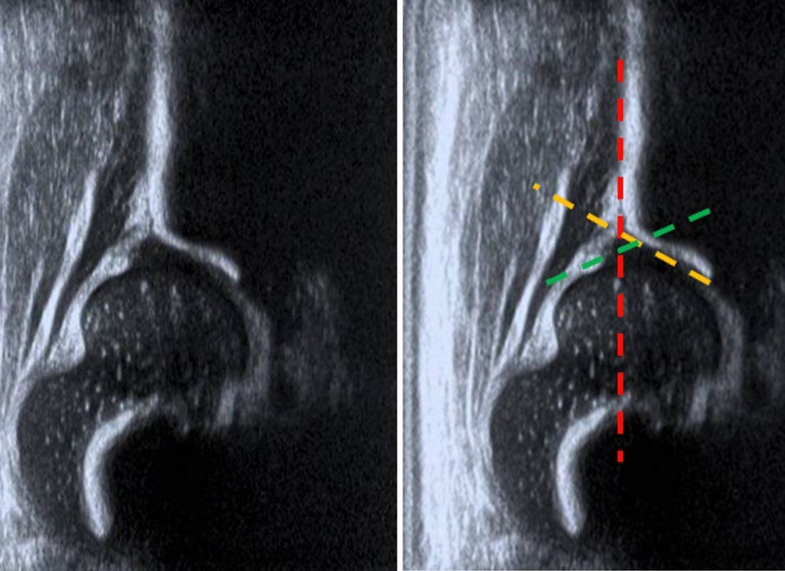
Considering both checklists and recognizing that both are useful for the study, lines will be drawn to result in the alpha and beta angles, thus determining the type of hip.
Check out ICODE's Application
The ICODE app has been developed to provide a reliable tool to assist health professionals in categorizing each of the hips screened using the Graf method.